4. < Back to the Campus

Welcome to 4. Flow Cytometry and Imaging

Using flow cytometry and fluorescent microscopy to confirm cells are making fluorescent proteins.

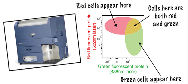
Aim: We have put our collagen plasmid into some mammalian cells. Now I need to check that my plasmid is in the cells by looking for the presence of the fluorescent proteins. Green fluorescent protein shines green and red fluorescent protein shines red when excited using the right wavelength of light.

I can use two methods to look at the protein levels in my cells.

To excite the cells means to give them energy by shining a laser at them (not to show them a fun time)

Flow Cytometry

I will use flow cytometry to look at the levels of protein in 1,000’s of cells. The machine excites the cells one by one using a laser and measures how green and red each cell is.
Each cell gets plotted on a graph as a single dot. The position on the graph indicates how much of that fluorescent protein there is in the cells plotted.

Fluorescence Microscopy

I will also look at the cells using fluorescence microscopy. I can excite the cells using the right wavelengths of light and see what colour they appear under a microscope. Unlike flow cytometry I can only look at a few cells at once, but I can see each cell lots in more detail.

Start Puzzle

Your time still start when you click the button

< Back to the Campus